Olivia Klee; Julia Buechler; Molly Fears ; Caroline Gosser; Jeffery Baker; Kahra Nix – This study describes a series of four cases where normal anatomy mimicked an abdominal aortic dissection
Olivia Klee; Julia Buechler; Molly Fears ; Caroline Gosser; Jeffery Baker; Kahra Nix – This study describes a series of four cases where normal anatomy mimicked an abdominal aortic dissection

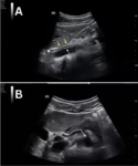
Olivia Klee; Julia Buechler; Molly Fears; Caroline Gosser; Kahra Nix – In this case series, we describe an artifact that mimics a dissection involving the abdominal aorta that was found on a young, healthy, thin female medical student who was acting as a standardized patient. A radiology-performed ultrasound of her abdomen confirmed the abdominal aorta as normal. This same artifact was subsequently seen on three additional young, healthy, thin, female medical students.
by Bill Ayach MD PhD; Aadil Dhansay MD1, Andrew Morris MD; James W. Tam MD; Davinder S. Jassal MD –
A 59 year old male presented with a 1 day history of non-exertional chest pain that was pleuritic in nature and aggravated by lying flat. His chest pain symptoms were preceded by a one week history of “flu-like” symptoms. Physical exam demonstrated a blood pressure of 114/55 mmHg, heart rate of 75 bpm, and a normal oxygen saturation on room air. Cardiac examination revealed a biphasic pericardial rub vs. to-and-fro murmur.