Archives
Figure-1-for-web

Simulator operation technique. (A) To operate the simulator, the participant places a reproduction of an ultrasound probe on a mannequin’s chest. (B) User interface. (C) Enlargement of three-dimensional visual guidance indicator. The position and orientation of the acquired image (blue) and anatomically correct image (green) are displayed with the angle error (white arc). (D) Enlargement of numeric report of skill metrics. The angle error is above and the probe placement error is below.
Album: test album
Categories: Image
Tags:
Figure 4 ECHO Abd Aortic Aneurysm Khidir-de-id
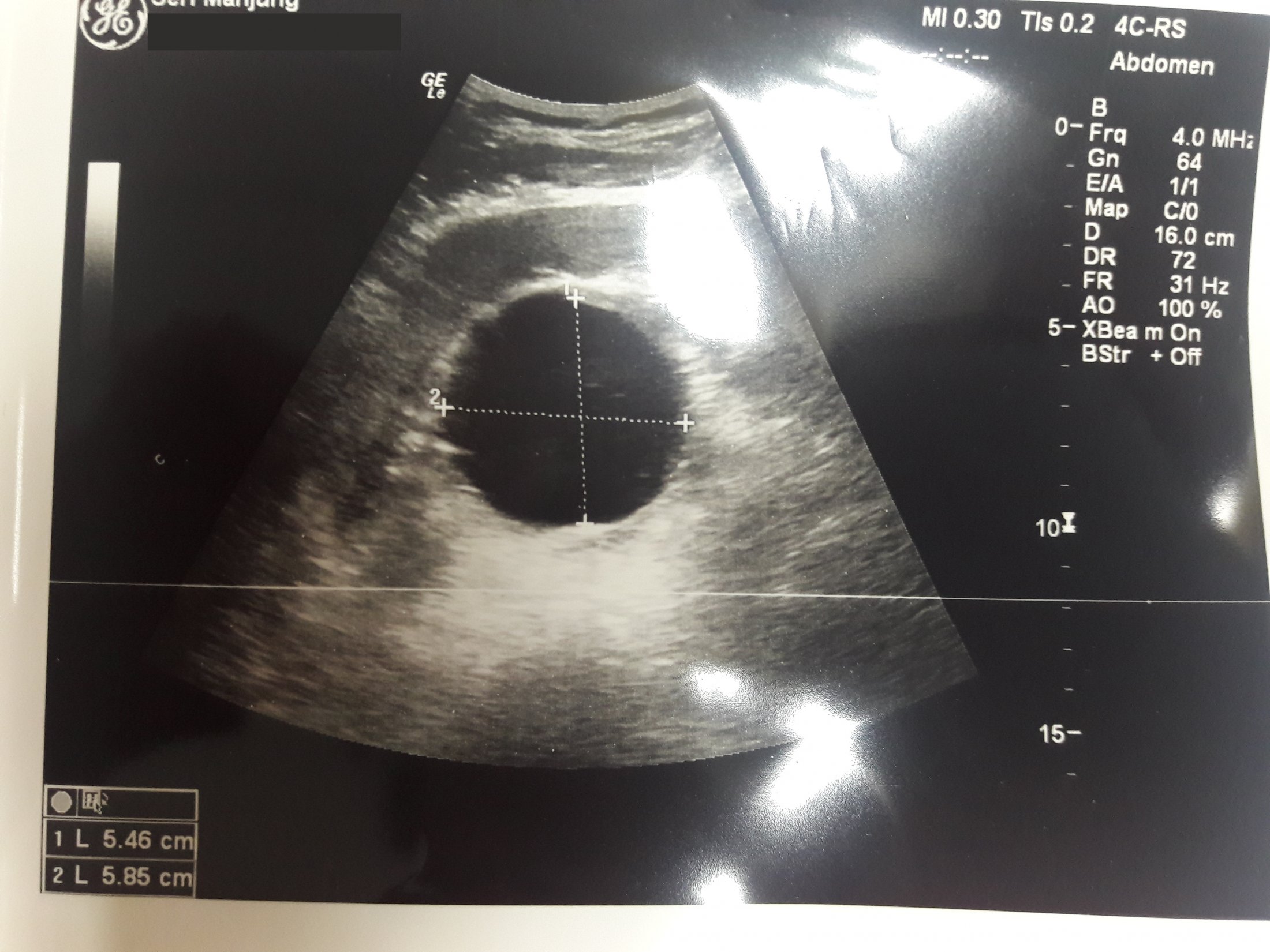
Abdominal aortic aneurysm measuring 5.4 x 5.8 cm with peri-aortic haematoma.
Album: test album
Categories: Image
Tags: