Categories: Thumbnail
Categories: Thumbnail
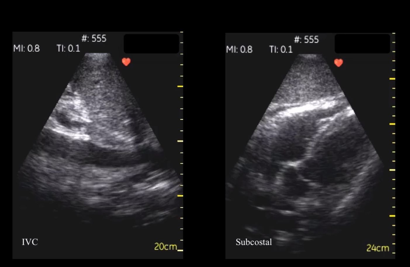
A 59 year old was admitted to the hospital and after a routine #POCUS was performed, an unexpected thin walled structure was observed in the abdominal cavity. The diagnosis can be found here: https://t.co/Ys1Kzd5mfK … #FOAMus #FOAMed #POCUSJournal @amerjohri pic.twitter.com/IUKOpbo4PE
— POCUS Journal (@POCUSJournal) April 2, 2019
Point of Care Ultrasound of Right Lower Abdominal Quadrant. Four still images captured from the 6 seconds of video recording.
Album: test album
Categories: Image
Tags:

Chest X-ray shows mediastinal widening suggestive of thoracic aortic dilatation.
Album: test album
Categories: Image
Tags:
Abdominal aortic aneurysm measuring 5.4 x 5.8 cm with peri-aortic haematoma.
Album: test album
Categories: Image
Tags:

Simulator operation technique. (A) To operate the simulator, the participant places a reproduction of an ultrasound probe on a mannequin’s chest. (B) User interface. (C) Enlargement of three-dimensional visual guidance indicator. The position and orientation of the acquired image (blue) and anatomically correct image (green) are displayed with the angle error (white arc). (D) Enlargement of numeric report of skill metrics. The angle error is above and the probe placement error is below.
Album: test album
Categories: Image
Tags: