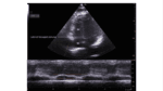
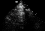
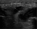
Yannis Amador – A 77-year-old man scheduled for coronary artery bypass grafting (CABG) experienced cardiac arrest immediately following the induction of anesthesia. His medical history was significant for hypertension, type II diabetes mellitus, and triple-vessel coronary artery disease with a proximal left main occlusion.
Issues: Vol 10 Iss 01 - APR 2025
POCUS Journal Volume 10, Issue 01, released April 2025
Perforation of the Interventricular Septum by a Temporary Transvenous Pacing Lead Detected by Cardiac Point of Care Ultrasound (POCUS)
Pablo Blanco; Liliana Figueroa – A 63-year-old woman was admitted with symptomatic complete atrioventricular block. Her medical history was notable for arterial hypertension, managed with enalapril. A TTP was successfully placed via the right internal jugular vein under the guidance of POCUS and intracavitary electrocardiogram (ECG), following this procedure:…
A Right Turn in Diagnosis: Highlighting the Importance of TAPSE in Isolated Right Ventricular Myocardial Infarction
Vasudha Dinesh; Arun A. Mohanan; Amaravathi Uthayakumar; Vinodha Chandrashekar – A 65-year-old man with a history of chronic tobacco and alcohol use but no known comorbidities presented to the emergency department with acute chest pain lasting three hours. The pain was central, tight, and radiated to the left arm, and was accompanied by profuse sweating.
Cardiac Point of Care Ultrasound (POCUS) Used to Diagnose Infective Endocarditis Following Multiple Negative Echocardiograms
Adriano Sanjuan; Daniel S Brenner; Heather Andrade; Alyson Bundy; Philip Clapham; Nathan Markus; Irina K Hariri; Edwin Jackson – We present a case of a 68-year-old African American man with a history of housing insecurity, chronic obstructive pulmonary disease, chronic hypoxic respiratory failure, heart failure with mildly reduced ejection fraction, chronic kidney disease, hypertension, opioid use disorder, schizophrenia, and mild mitral, tricuspid, and aortic valve insufficiency.
Health System Impact of Emergency Department-based Vascular Access Program in Patients with Difficult Intravenous Access
Nathan P. Roll; Shilpa Raju; Micah Ownbey; Jamal Jones; Christy Hopkins; Jennifer Cotton – We describe the impact of an Emergency Department (ED) based vascular access program with a specialized team of paramedics and emergency medical technicians (EMTs). This team is trained in USG PIV access to assist with patients that have difficult intravenous access (DIVA) both in the ED and throughout the academic medical center.
The Sound of Safety: DIVOT (Doppler Imaging for Vascular Orientation in Thoracic Procedures) Protocol
Amy Fraser; Daniel S. Brenner; Matthew Coghlan;Heather Andrade; Maya Haouili; William Graham Carlos; Edwin Jackson – This protocol provides comprehensive procedural guidance to minimize the risk of hemorrhagic complications associated with thoracentesis and percutaneous chest tube placement, supporting our commitment to eliminating preventable harm.
A Point of Care Ultrasound (POCUS) Artifact Mimicking an Aortic Dissection: A Case Series
Olivia Klee; Julia Buechler; Molly Fears; Caroline Gosser; Kahra Nix – In this case series, we describe an artifact that mimics a dissection involving the abdominal aorta that was found on a young, healthy, thin female medical student who was acting as a standardized patient. A radiology-performed ultrasound of her abdomen confirmed the abdominal aorta as normal. This same artifact was subsequently seen on three additional young, healthy, thin, female medical students.
Interrater Agreement of Physicians Identifying Lung Sliding Artifact on B-Mode And M-Mode Point of Care Ultrasound (POCUS)
Ross Prager; Hans Clausdorff Fiedler; Delaney Smith; Derek Wu; Robert Arntfield – Chest point of care ultrasound (POCUS) is a first-line diagnostic test to identify lung sliding, an important artifact to diagnose or rule out pneumothorax. Despite enthusiastic adoption of this modality, the interrater reliability for physicians to identify lung sliding is unknown.
Rib Reverberation: An Important New Artifact in Lung Ultrasound
Yulei Cao; Jennifer K. Sun; Cameron M. Baston – We present a case report describing horizontal sonographic artifacts that resemble A-lines generated by a rib.
Parotid Sialolithiasis Diagnosed on Point of Care Ultrasound (POCUS)
Heather Lystad; Elaine Yu; Rachna Subramony – A 58-year-old woman with a past medical history of irritable bowel syndrome presented to the emergency department due to gradual swelling and pain on the left side of her face for three weeks. She also reported feeling a lump on her inner cheek. The patient experienced worsened swelling, pain with opening her mouth, and shooting pains on the left side of her face. She denied any fevers or chills.


